


El efecto "puerta de pantalla": Por qué los fibroscopios de 3,5 mm están anclados en la década de 1990


En una era de pantallas quirúrgicas de ultra alta definición y precisión quirúrgica, la vista a través de un endoscopio de fibra óptica estándar de 3,5 mm a menudo todavía parece una transmisión de baja resolución de 1995. La limitación fundamental del endoscopio de fibra óptica de 3,5 mm radica en la pixelación inherente a su construcción. Debido a que un diámetro exterior de 3,5 mm también debe acomodar un canal de trabajo de 5 Fr (1,67 mm), el espacio restante para la óptica se restringe a una media luna estrecha e irregular. Para llenar este pequeño espacio, los fabricantes confían en haces de fibra óptica compuestos por miles de hilos de vidrio individuales. Cada hilo actúa como un solo píxel, creando un patrón de panal donde el revestimiento negro entre las fibras es visible para el cirujano. Esto se conoce como el efecto de puerta de pantalla, y obliga al ojo humano a filtrar constantemente una malla cuadriculada para ver el tejido subyacente. Al igual que un televisor de tubo de los años 90, la resolución está limitada por el número físico de fibras que se pueden empaquetar en el espacio óptico limitado.
Más allá de la granularidad visual inmediata, los sistemas de fibra óptica enfrentan una crisis de durabilidad que degrada aún más la imagen con el tiempo. Debido a que estos endoscopios están construidos con miles de delicados hilos de vidrio, son altamente susceptibles a la rotura de fibras o fracturas del núcleo durante el manejo o la esterilización rutinarios. Cada vez que una sola fibra se rompe, aparece un punto negro permanente en el campo de visión. A diferencia de los sistemas de lentes sólidos, no hay forma de reparar estos píxeles muertos. La imagen simplemente se vuelve más desordenada y obstruida a medida que el endoscopio envejece. Esta degradación es la razón por la cual un endoscopio de fibra óptica puede parecer aceptable recién salido de la caja, pero rápidamente retrocede a una vista de baja fidelidad y mapeada por píxeles que oscurece las patologías finas requeridas para la navegación quirúrgica de alto riesgo.
En última instancia, el problema de la fibra óptica es un techo físico. Para mantener el diámetro de 3,5 mm y al mismo tiempo un canal de trabajo, los fabricantes deben usar estos pequeños hilos de vidrio, pero esos hilos no pueden transmitir la trayectoria de luz continua y cruda necesaria para la visualización moderna de alta definición. El cirujano esencialmente está viendo una aproximación digital de un sitio quirúrgico en lugar de una reproducción analógica clara. Esto resulta en una pérdida significativa de conciencia espacial y precisión del color, lo que convierte al sistema de fibra óptica en un cuello de botella para procedimientos donde la integridad visual es el requisito principal para la seguridad del paciente y la velocidad operatoria.
Para proporcionar respaldo científico a la analogía de la televisión de los años 90, los investigadores han identificado varios conceptos técnicos y fuentes creíbles que explican la pixelación y los problemas de durabilidad inherentes a los haces de fibra óptica en endoscopios de pequeño diámetro. Un estudio histórico publicado en el Journal of Biophotonics señala que cuando los diámetros de los endoscopios se reducen al rango de 1 mm a 3 mm, los haces de fibra óptica tradicionales proporcionan una resolución tan pobre que la agudeza visual a menudo cumple con la definición legal de ceguera. Mientras que un monitor quirúrgico HD moderno muestra 1920 píxeles de ancho, un haz estándar de 30.000 fibras solo ofrece aproximadamente 250 píxeles en todo el campo de visión.
El aspecto de puerta de pantalla es un artefacto documentado en ingeniería óptica. Cada fibra de vidrio en un endoscopio debe estar rodeada por una capa de revestimiento con un índice de refracción más bajo para evitar que la luz se escape a través de la reflexión interna total. Este revestimiento es opaco y ocupa un porcentaje significativo del espacio óptico disponible en un endoscopio pequeño de 3,5 mm con un canal de trabajo. Este espacio no visual crea la rejilla oscura en forma de panal que obstruye la vista del cirujano, un fenómeno explorado en el Journal of Clinical Medicine con respecto a los algoritmos de eliminación de panal y mejora de la imagen.
A diferencia de los sistemas de lentes de varilla, que son cilindros de vidrio sólidos, los haces de fibra están hechos de hilos de vidrio fusionados que son susceptibles a golpes mecánicos y térmicos durante el estrés de la sala de operaciones y el calor del autoclave. Doblar o dejar caer un endoscopio de fibra provoca una fractura del núcleo. Cada núcleo fracturado deja de transmitir luz, lo que resulta en un píxel muerto negro permanente. Un estudio en Medical Devices: Evidence and Research encontró que la transmisión de luz de fibra óptica disminuye significativamente antes en el ciclo de vida del endoscopio en comparación con los sistemas de varilla debido a estas microfracturas.
Además, la Sociedad Internacional de Óptica y Fotónica (SPIE) explica que el brillo y la claridad de un endoscopio están determinados por su eficiencia de recolección de luz. En un endoscopio de 3,5 mm con un canal de 5 Fr, la apertura es tan pequeña que el haz de fibra no puede capturar suficiente luz para competir con un sistema de lentes de varilla. El sistema de lentes de varilla elimina los espacios de aire de las lentes más antiguas, lo que permite un índice de refracción mucho más alto y una imagen analógica continua que no depende de hilos pixelados.
Contexto científico sobre la agudeza: Examine la investigación sobre la endoscopia de fibra de barrido y las limitaciones de los haces de fibra coherente tradicionales en catéteres pequeños: Journal of Biophotonics: Wide-field Full-color Imaging.
Explore artefactos ópticos: Comprenda la ingeniería detrás del aspecto de "puerta de pantalla" y los intentos digitales de mejorar las imágenes de panal: Journal of Clinical Medicine: Honeycomb-Removal Algorithms.
Analice la degradación del ciclo de vida: Revise los datos de 2025 que evalúan cómo las microfracturas en la fibra óptica provocan una falla óptica temprana: Medical Devices: Evaluation of Optical Degradation Patterns.
Domine la física: Acceda al texto fundamental sobre óptica de endoscopios y la eficiencia de recolección de luz de los sistemas de lentes de varilla: SPIE Press: Endoscope Optics.
Referencias:
Lee, M. C., et al. (2010). "Scanning fiber endoscopy with highly flexible, 1-mm catheterscopes for wide-field, full-color imaging." Journal of Biophotonics.
Journal of Clinical Medicine (2022). "Effects of Image Processing Using Honeycomb-Removal and Image-Sharpening Algorithms."
Mierlo, S., et al. (2025). "Evaluation of Optical Degradation Patterns in Rigid Endoscopes." Medical Devices: Evidence and Research.
Capítulo 8. "Endoscope Optics." SPIE Press. Sociedad Internacional de Óptica y Fotónica.
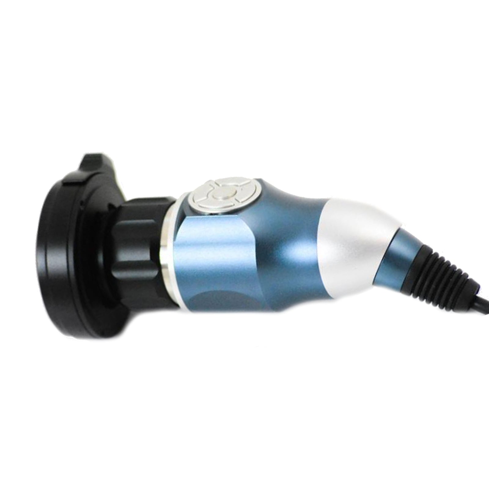
Experimente una forma más inteligente de adquirir equipos de endoscopia, todo en un solo lugar.
✔ Amplia selección de marcas de endoscopia de confianza
✔ Precios competitivos sin comprometer la calidad
✔ Acceso rápido y fiable a las herramientas que necesita, cuando las necesita
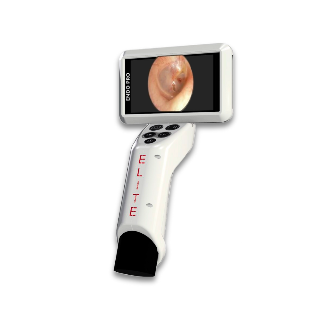
👉 Descubra cómo EndoPro® 3D puede transformar su técnica quirúrgica.

Disclaimer: All product names, logos, brands, and trademarks displayed on this website are the property of their respective owners and are used solely for informational and descriptive purposes. The appearance of any third-party trademarks does not imply any affiliation, endorsement, sponsorship, or authorization by the trademark holder. This ecommerce store and its product listings operate independently and are not affiliated with, authorized by, endorsed by, or officially connected to KARL STORZ, Medtronic, Olympus Corporation, Richard Wolf GmbH, Stryker Corporation, or any other trademark owners referenced on this site, including their respective subsidiaries or affiliates.